
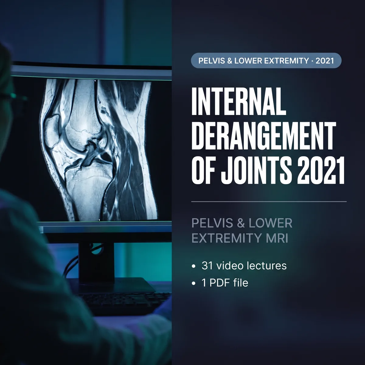
This course is an update on MR imaging in the assessment of musculoskeletal disorders of the pelvis, hip, knee, ankle and foot.
Essential anatomy, physiology and pathology are emphasized to explain imaging findings, and MR findings in common lower-extremity problems are compared with those of other imaging methods. Donald Resnick leads a faculty of musculoskeletal radiologists.
What’s included
- 31 video lectures · 1 PDF
- On-demand streaming access to the recorded sessions
- Self-study material; does not include CME or MOC credit.
What you’ll learn
- Assess MR images in patients with internal derangements of peripheral joints
- Describe the anatomic features fundamental to accurate interpretation of these MR findings
- Formulate a reasonable differential diagnosis and identify the most likely diagnosis
- Understand the pathogenesis and imaging of common and important disorders of the pelvis and lower extremities
Course contents
Joints, bone and muscle
- Articular Disorders of Synovium-Lined Joints: Morphology, Inflammatory and Degenerative Disorders, Tumors and Tumor-Like Disorders
- Focus Session: Osteonecrosis Versus Insufficiency Fractures, with Emphasis on the Hip and Knee
- Chondral, Osteochondral and Subchondral Injuries: Anatomy, Pathophysiology, Terminology and MR Imaging
- Cartilage Imaging: Routine and Advanced Methods
- Stress-Related Abnormalities of the Skeleton with Emphasis on the Pelvis and Lower Extremity
- Focus Session: Muscle Disorders: Anatomy, Pathophysiology and General Abnormalities
- Focus Session: Bone Marrow: Normal and Abnormal, with Emphasis on MRI
- Focus Session: Important Entrapment Neuropathies of the Pelvis and Lower Extremity
Pelvis and hip
- Muscles and Tendons About the Pelvis and Hip: Anatomy, Strains and Tears
- Labral Abnormalities and External and Internal Femoroacetabular Impingement
Knee
- Meniscus: Structure, Function and Patterns of Failure
- Discoid Menisci and Other Anomalies
- Knee Ligaments: Anatomy, Biomechanics and Footprints of Injury
- Anterior Cruciate Ligament
- Posterior Cruciate Ligament
- Medial Supporting Structures of the Knee
- Lateral Supporting Structures of the Knee
- Postoperative Ligaments with Emphasis on the Anterior Cruciate Ligament
- Patellofemoral Maltracking and Patellar Instability/Dislocation
- Quadriceps and Patellar Tendons, Fat Pads, Bursae and Plicae
- Popliteal Fossa
- Focus Session: MRI/Arthroscopy Correlation
Ankle and foot
- Tendons: Normal Anatomy
- Tendons: Tendinosis, Tenosynovitis, Tendon Tears and Other Abnormalities
- Ligaments: Normal Anatomy
- Ligaments: Patterns of Injury
- Adult Acquired Flatfoot Deformity
- Fractures and Dislocations of the Ankle and Foot: Role of CT Scanning
- Tumors and Tumor-Like Lesions of the Ankle and Foot
- Osteomyelitis, Septic Arthritis and Soft Tissue Infection with Emphasis on the Diabetic Foot
- Rapid-Fire Case Review: Tarsal Coalition, Osteochondritis Dissecans of the Talus, Metatarsalgia, Plantar Aponeurosis
Faculty
- Donald L. Resnick, Christine B. Chung, Mini N. Pathria, Evelyne A. Fliszar, Brady Huang, Lucas Hiller, Eric Y. Chang, Karen C. Chen, Tudor Hughes, Edward Smitaman
Who should attend
- Practicing radiologists
- Orthopaedic surgeons, rheumatologists, podiatrists and sports medicine physicians




Reviews
There are no reviews yet