
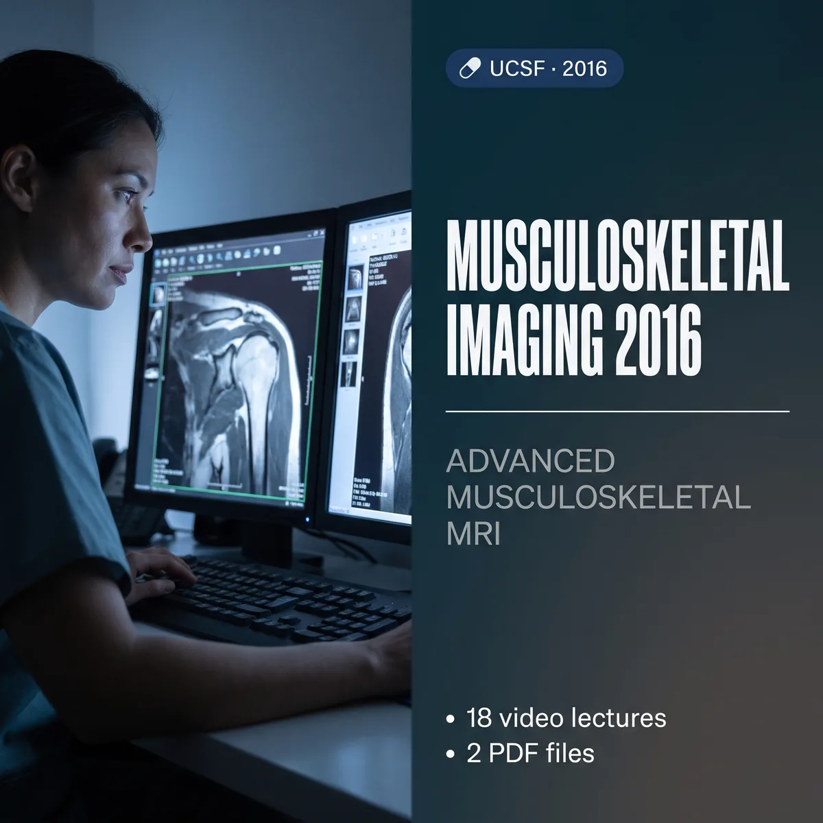
UCSF Musculoskeletal Imaging 2016 (released May 2016) was planned for radiologists, orthopaedic surgeons, sports medicine physicians and MRI technologists with basic or intermediate knowledge of musculoskeletal MRI who want to reach a more advanced level.
The 18 lectures cover the shoulder, wrist, hip, knee, ankle, fingers and toes, bone marrow and cartilage, bone tumors, cervical spine trauma, peripheral nerves and imaging after arthroplasty.
What’s included
- 18 video lectures · syllabus and question PDFs
- On-demand streaming access to the recorded sessions
- Self-study material; does not include CME or MOC credit.
What you’ll learn
- Relate MR findings of the shoulder and knee to clinically relevant orthopaedic disorders
- Develop optimized musculoskeletal protocols, including metal-suppression protocols after joint replacement
- Understand the MRI anatomy of the shoulder, wrist, hip, knee, ankle, fingers and toes
- Identify abnormalities of the joints, muscles, bone marrow and cartilage and their clinical significance
- Recognize common bone tumors with a pertinent differential diagnosis
- Recognize abnormalities related to cervical spine trauma
- Understand peripheral nerve imaging protocols and findings
Course contents
- MRI of the Postoperative Shoulder
- Digital MRI: The Fingers and Toes
- Pitfalls in Knee MRI
- MRI Following Joint Arthroplasty
- Pseudotumors
- Imaging of Cervical Spine Trauma
- Pitfalls in Shoulder MRI
- Cartilage and Osteochondral Disease
- MRI of the Hip Labrum and FAI
- MRI of the Hip Tendons
- MRI of the Wrist: Ligaments and TFCC
- Patterns of Bone Marrow Edema
- Rotator Cuff and Impingement
- Knee ACL
- Knee Meniscus
- Ankle MRI
- Peripheral Nerve Imaging
- Bone Tumors
Faculty
- Lynne S. Steinbach, John F. Feller, Daria Motamedi, Thomas M. Link, Christine B. Chung
Who should attend
- Diagnostic radiologists
- Orthopaedic surgeons, sports medicine physicians and MRI technologists




Reviews
There are no reviews yet